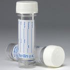
Cerebrospinal Fluid (CSF) Non-Gynae Cytology
The cerebrospinal fluid (CSF) is produced from arterial blood by the choroid plexuses of the lateral and fourth ventricles by a combined process of diffusion, pinocytosis and active transfer. A small amount is also produced by ependymal cells.
Non-Gynae Cytopathology
The Functions of the CSF
- Buoyancy effect: Because brain weight is effectively reduced by more than 95%, shearing and tearing forces on neural tissue are greatly minimized.
- Intracranial volume adjustment CSF volume can be adjusted, increasing or decreasing acutely in response to blood volume changes or chronically in response to tissue atrophy or tumour growth.
- Micronutrient transport: Nucleosides, pyrimidines, vitamin C, and other nutrients are transported by the choroid plexus to CSF and eventually to brain cells.
- Protein and peptide supply Macromolecules like transthyretin, insulin-like growth factor, and thyroxine are transported by the choroid plexus into CSF for carriage to target cells in brain.
- Buffer reservoir: When brain interstitial tissue fluid concentrations of H+, K+, and glucose are altered, the ventricular fluid can help to buffer the extracellular fluid changes.
- Sink or drainage action: Metabolites of neurotransmitters, and protein products of catabolism or tissue breakdown are cleared from the CNS by active transporters in the choroid plexus or by bulk CSF drainage pathways to venous blood and the lymphatics.
- Immune system mediation: Cells adjacent to ventricles have antigen-presenting capabilities. Some CSF proteins drain into cervical lymphatics, with the potential for inducing antibody reactions.
- Information transfer: Neurotransmitter agents like amino acids and peptides may be transported by CSF over distances to bind to receptors in the parasynaptic mode.
- Drug delivery: Some drugs do not readily cross the blood-brain barrier but can be transported into the CSF by endogenous proteins in choroid plexus epithelial membranes.
- Normal CSF characteristics: Crystal clear, opening pressure of 80-180 mmH2O (in patients with high BMI slightly higher pressures can be normal). Up to 5 mononuclear cells, 0-5 red blood cells, Glucose level 60-70% of plasma values, protein =<0.4 g/L, lower in the ventricles.
Abnormalities in CSF Pressure
- Hydrocephalus:
- Communicating: Impairment of CSF absorption (e.g. blockage of arachnoid villi after a subarachnoid haemorrhage).
- Non-Communicating: Obstruction of CSF circulation (e.g. Fourth ventricle tumour)
- “Normal Pressure Hydrocephalus” which mainly occurs in the elderly with mental confusion, gait abnormalities and urinary incontinence, is a misnomer as plateaus of high pressure have been demonstrated despite “normal” spot readings.
- Idiopathic Intracranial Hypertension: Occurs predominantly in overweight young females presenting with headaches, and visual loss. Optic nerves are at risk without treatment. Similar presentation can occur following taking tetracyclines, Vitamin A supplements.
- Space occupying lesions/cerebral oedema/haemorrhage: Leading to a general increase in CSF pressure.
- Dural venous sinus thrombosis: Impairment of CSF drainage.
- CSF hypotension: Iatrogenic (e.g. after lumbar puncture) or spontaneous, due to dural leak.
Abnormalities in CSF constituents
- Elevated White blood cells: Infection (meningitis,encephalitis)-Neutrophils prevalent in bacterial meningitis, Lymphocytes prevalent in viral and tuberculous meningitis, Reactive to neoplastic involvement, Autoimmune disorders, Sarcoidosis.
- Elevated Red blood cells: Traumatic tap, Subarachnoid haemorrhage; Xanthochromia screen looking at the RBC breakdown products (bilirubin and oxyhaemoglobin) can be diagnostic.
- Low Glucose: Hypoglycaemia; Bacterial, Fungal, or Tuberculous meningitis, Mumps and lymphocytic choriomeningitis infections (otherwise unusual in viral meningitis), Subarachnoid haemorrhage, Sarcoidosis.
- Elevated protein: Inflammatory conditions, Infection, CSF block, Neoplasms.
- Neoplastic cells: Primary or metastatic CNS involvement, up to 3 attempts at lumbar puncture might be required to detect the abnormal cells.
- Oligoclonal bands: CNS demyelination.
- Specific tests: Positive TPPA in syphilis, Positive 14-3-3 protein in Sporadic or Variant CJD, Positive Viral PCR in meningitis.
Other Useful Information and Links:-:-
- Cerebrospinal Fluid an Overview
- Examination of the Cerebrospinal Fluid: Lumbar Puncture
- CSF Xanthochromia Testing Guidelines
- CSF Examination Document (CSF-1) Form
Notes
- A CSF non-gynae cytology report is based on cellular morphology leading to an assessment of malignant potential. Non-gynae Cytology investigation of CSF may be of use in patients with the following clinical presentations
- Diagnosis and monitoring of haematological malignancies with CNS involvement
- ? Disseminated malignancy leading to neurological symptoms
- ? CNS tumour
Sample Requirements

- Whilst the largest possible volume is most likely to yield meaningful non-gynae cytology this must be prioritised against other investigations in microbiology and chemistry. CSF samples from patients with suspected meningitis must be sent for microbiological investigation, which includes a white cell and red cell count and where relevant, a white cell differential.
Storage/Transport
- CSF is a poor cell preservation medium. To maximise diagnostic accuracy it is essential that specimens are transported to the laboratory urgently. The CSF transport flowchart has been agreed to minimise the risk of problems in this area.
- Please note that failure to follow this pathway will result in an ACI being documented.
Required Information
All samples must be accompanied by a yellow non gynae cytology request form. The completion of the form and the labelling of the sample must conform to the Policy for Specimen and Request Form Labelling for Pathology Users.
Turnaround Time
Urgent CSF samples will be reported within the same working day when received in the laboratory by 3.00pm.
For a sample to be treated as urgent the laboratory must be notified on Cheltenham telephone number 0300 422 4219.